The patient is a 59 year old man with a new diagnosis of interstitial lung disease. A chest CT scan identified a 2.3 cm solid right upper lobe mass with calcifications. Presumed granulomata were also identified. He is a non-smoker and denies weight loss, malaise, or productive cough. CT-guided fine needle aspiration was performed.
(Click each image to enlarge)
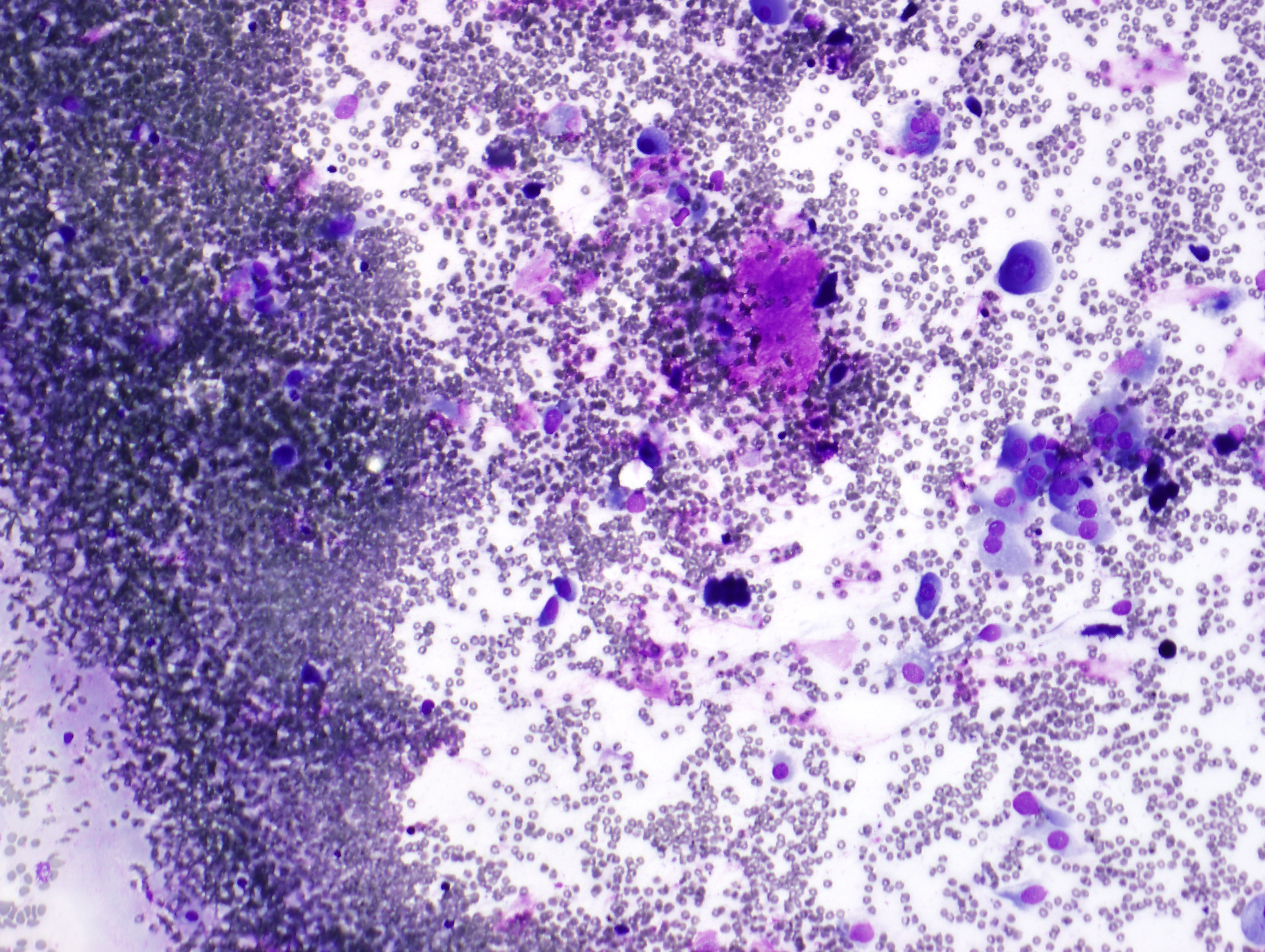
| FNA of lung lesion, Diff Quik at 10x | |
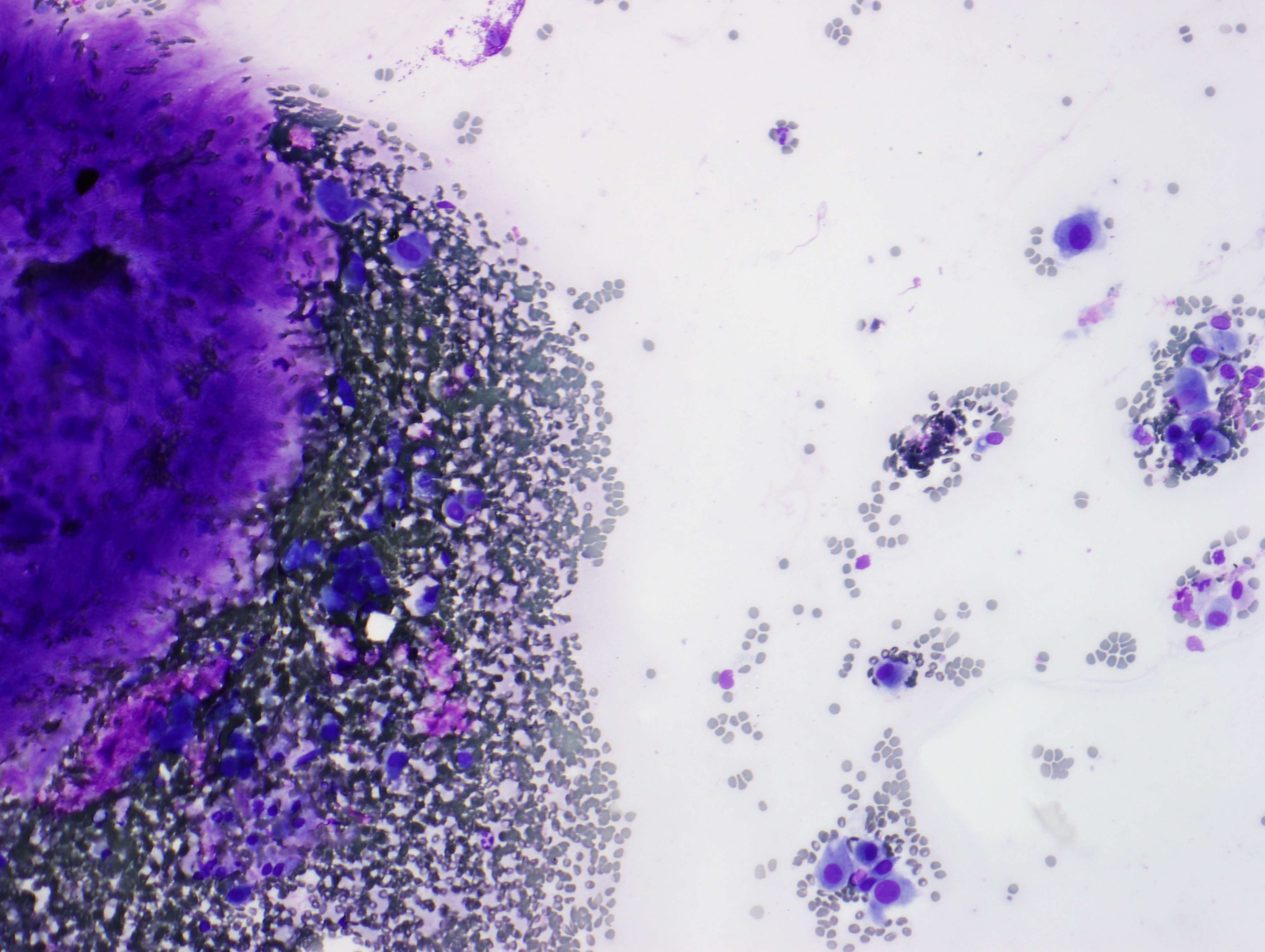
| FNA of lung lesion, Diff Quik at 10x | |
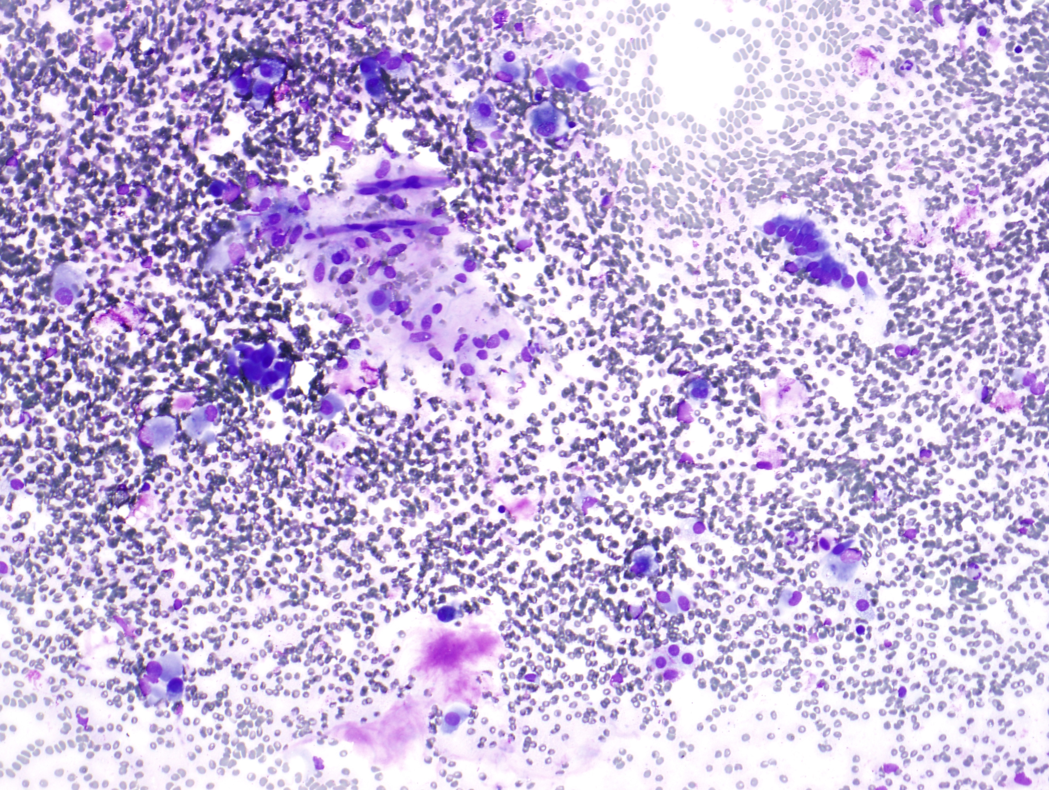
| FNA of lung lesion, Diff Quik at 10x |