
The patient is a 54 year old white female with a history of triple negative breast cancer. BRCA testing was negative. Routine Pap test identified the presence of atypical glandular cells (AGC). Concurrent HPV results were negative. As follow-up, her CA125 Ser QN was 16.5 units/ml. A pelvic ultrasound revealed a calcified mass on her left ovary.
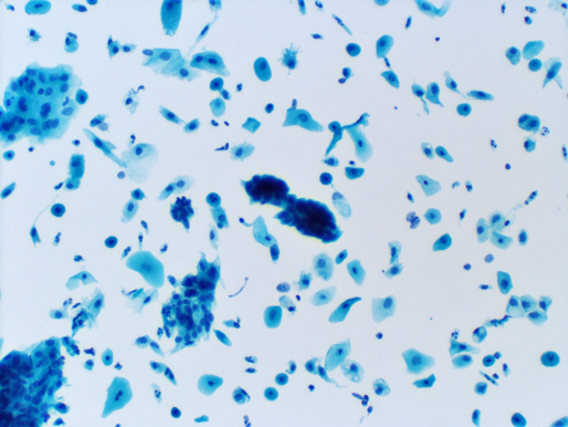
Figure 1 Title: Liquid based Pap test with Papanicolaou stain featuring hyperchromatic crowded groups, 100X
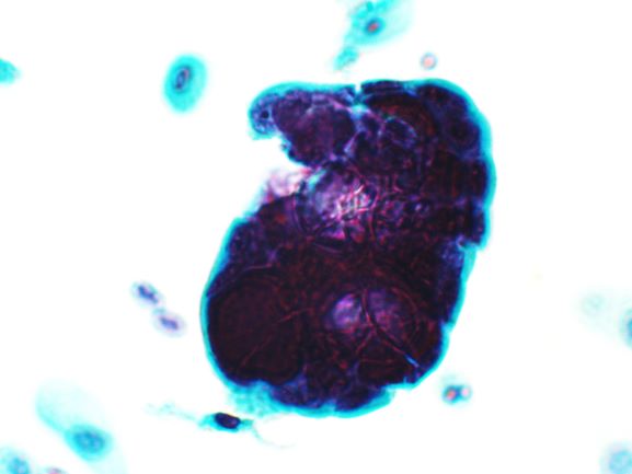
Figure 2 Title: Liquid based Pap test, Papanicolaou stain featuring hyperchromatic ball like grouping, 400X
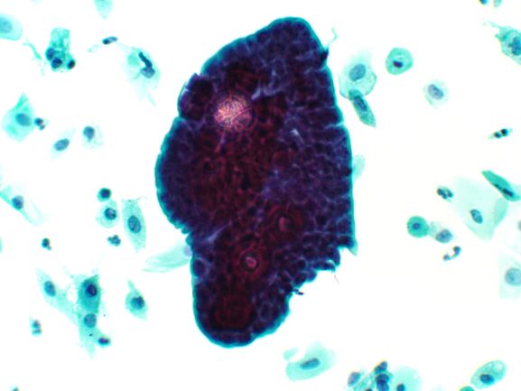
Figure 3 Title: Liquid based Pap test, Papanicolaou stain featuring a hyperchromatic, three dimensional group with a possible psammoma body, 200X
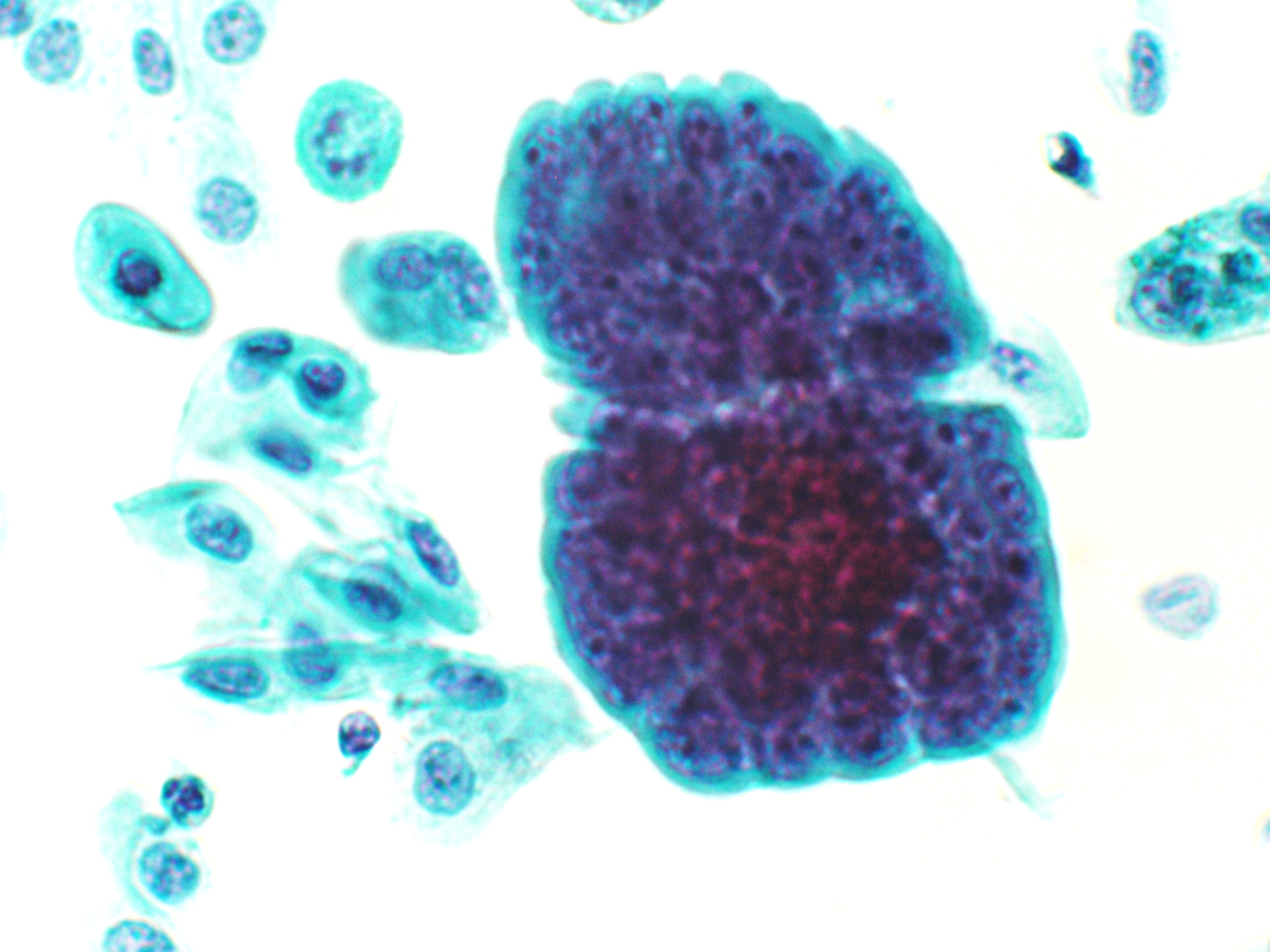
Figure 4 Title: Liquid based Pap test featuring a large, three-dimensional, hyperchromatic group, 400X