42 year old male presented with a 7.0 cm large right pelvic soft tissue mass. [Figures 1-2] An image-guided fine needle aspiration [Figures 3-5] and core biopsy [Figures 6-10] were performed and evaluated. Confirmatory immunohistochemical studies were performed on the core biopsy [Figures 8-10], and demonstrated positivity for vimentin, CD99 (O13), FLI-1, and AE1/3 (focal), while the following immunostains were negative: CD45, CD20, CD30, CD34, CD57, Myogenin, S100, and Desmin.
Figure 1: 7.0 cm intramuscular mass in anterior right pelvic soft tissue. MRI studies.
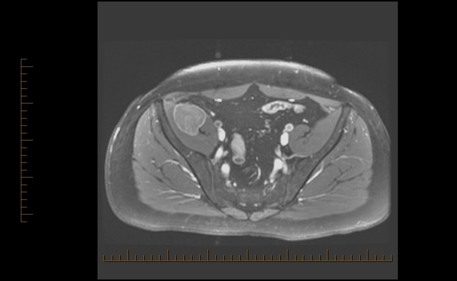
Figure 2: Fine needle aspiration guided biopsy of the mass. CT scan of pelvis.
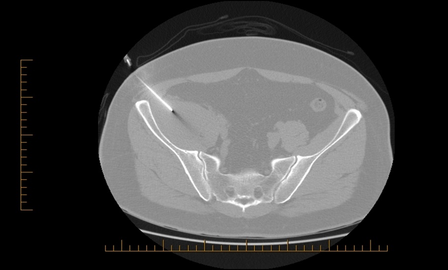
Figure 3: Malignant round cell tumor. Cytospin, Papanicolaou stain, 100 x magnification.
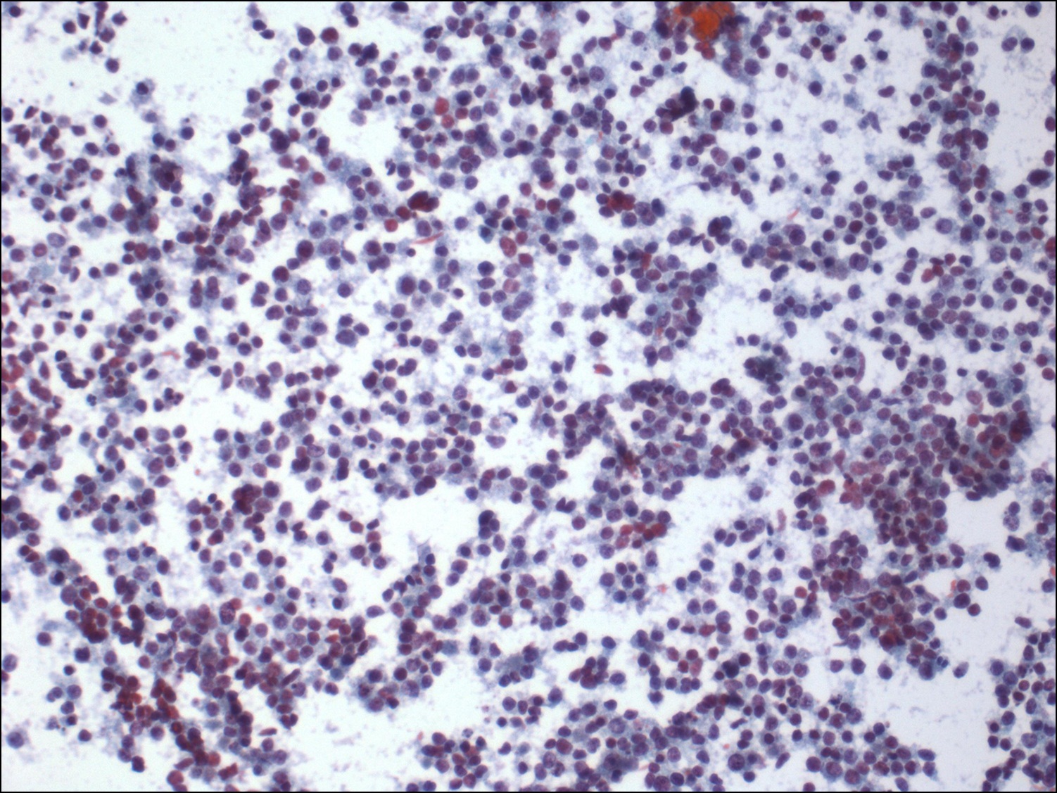
Figure 4: Malignant round cell tumor. Cytospin, Papanicolaou stain, 400 x magnification.
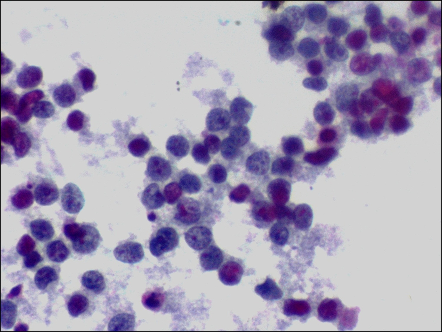
Figure 5: Malignant round cell tumor. Cell block, H&E stain, 100 x magnification.
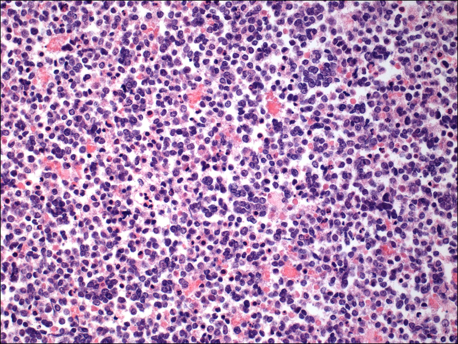
Figure 6: Malignant round cell tumor favors Ewing’s/PNET. Homer Wright rosettes are readily identified. Core biopsy, H&E stain, 100 x magnification.
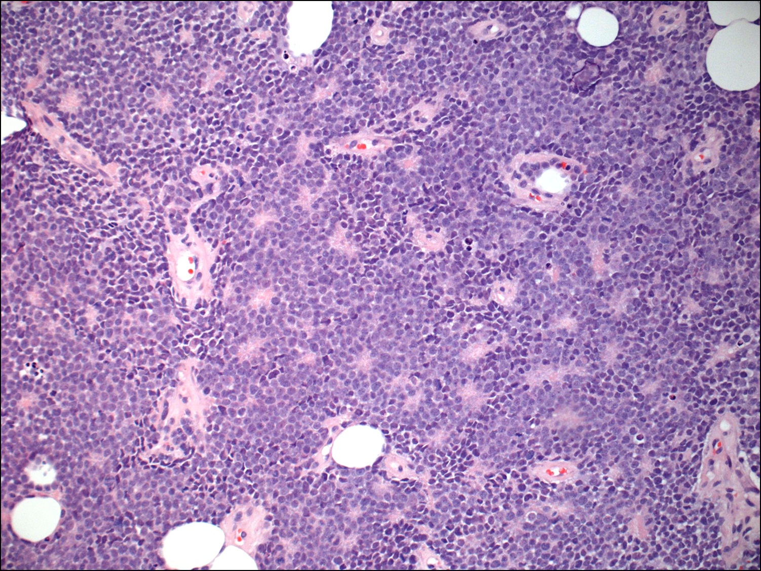
Figure 7: Malignant round cell tumor favors Ewing’s/PNET. Core biopsy, H&E stain, 400 x magnification.
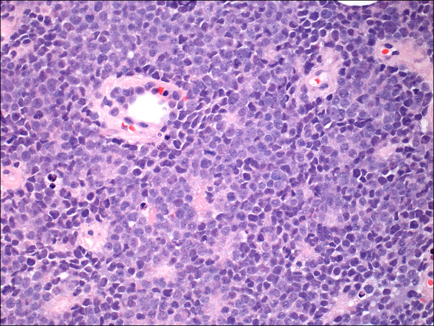
Figure 8: CD99 (O13) immunostain: Positive membranous expression. Core biopsy, 100 x magnification.
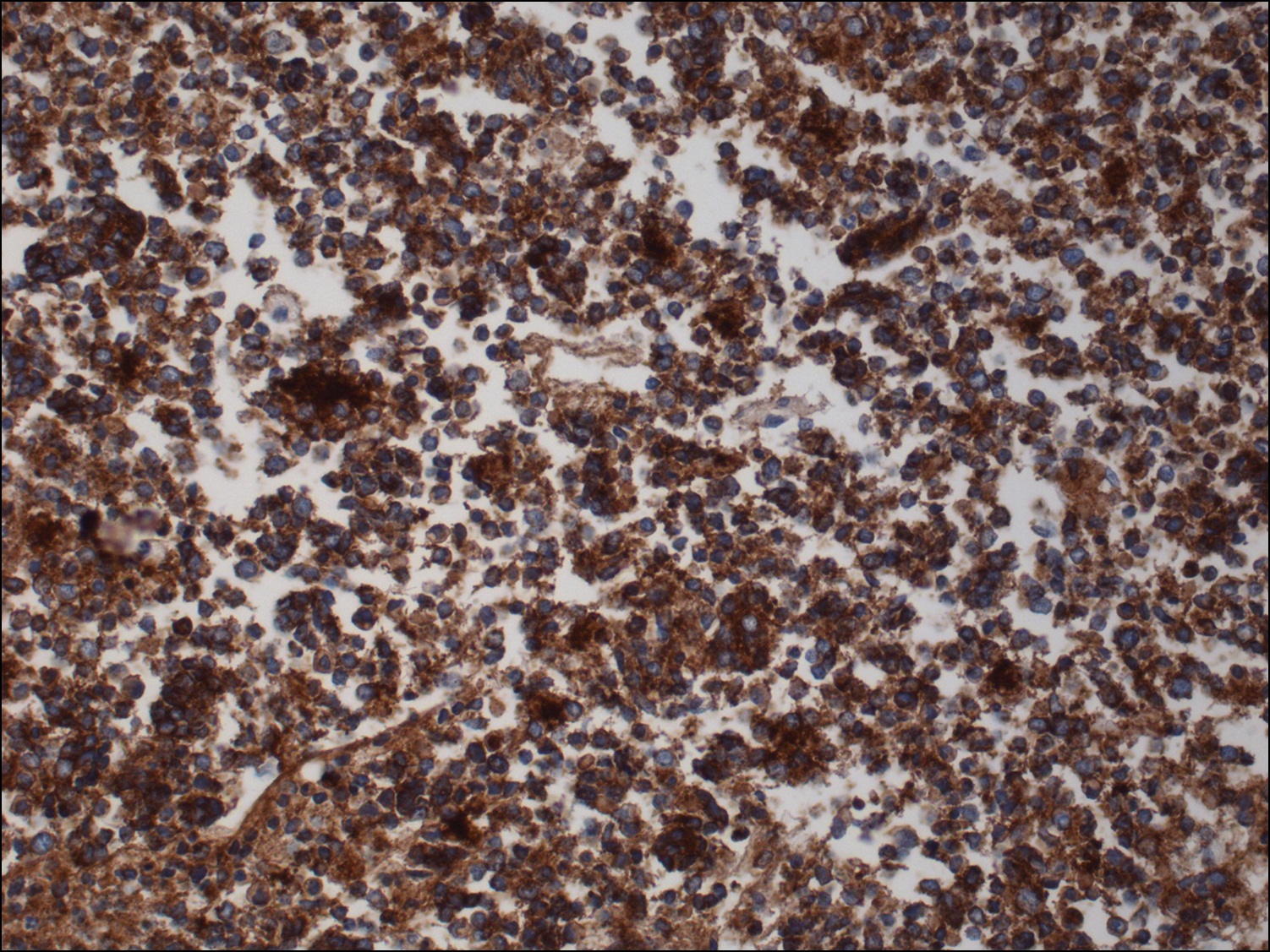
Figure 9: FLI-1 immunostain: Positive nuclear expression. Core biopsy, 100 x magnification.
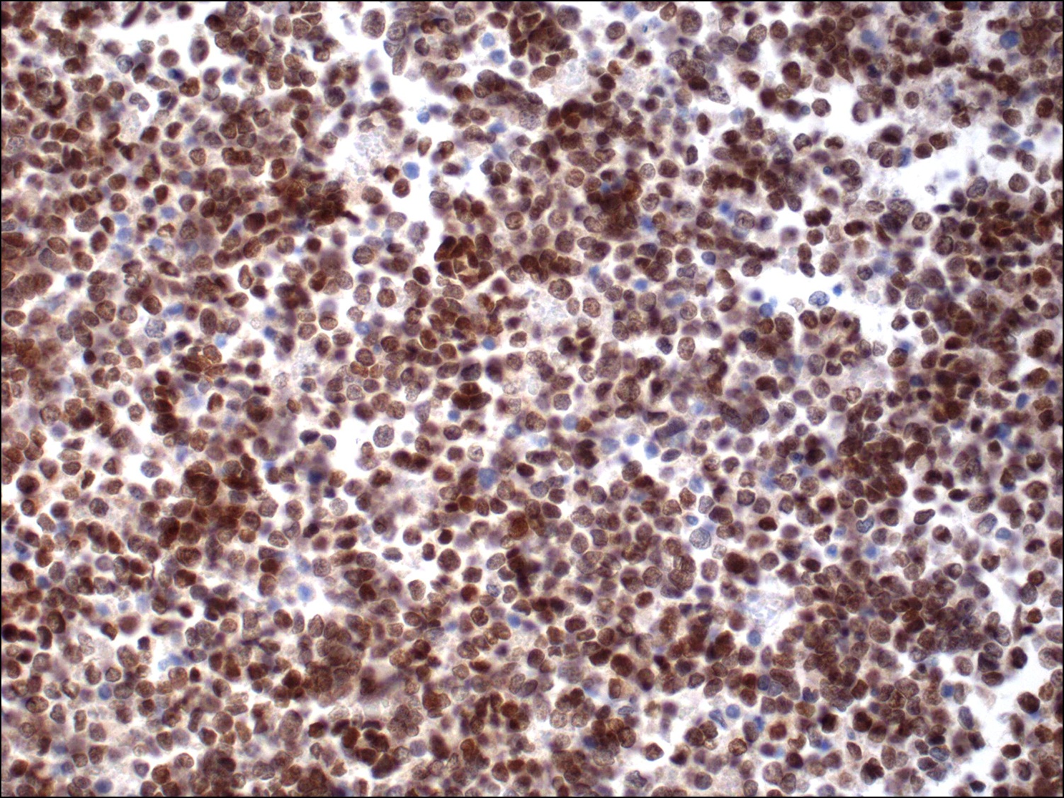
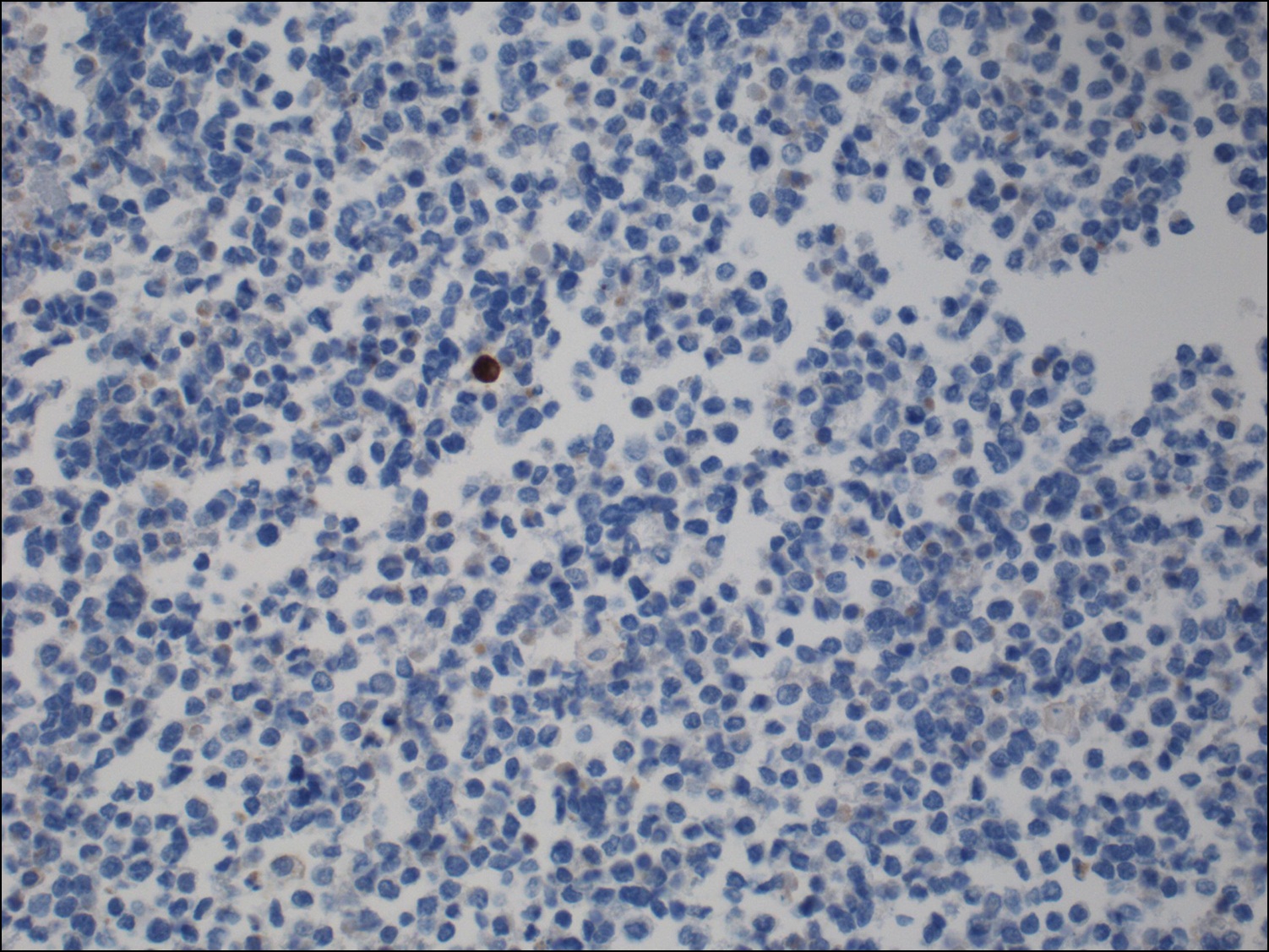
Figure 10: CD45 (LCA) immunostain: negative expression. Core biopsy, 100 x magnification.