
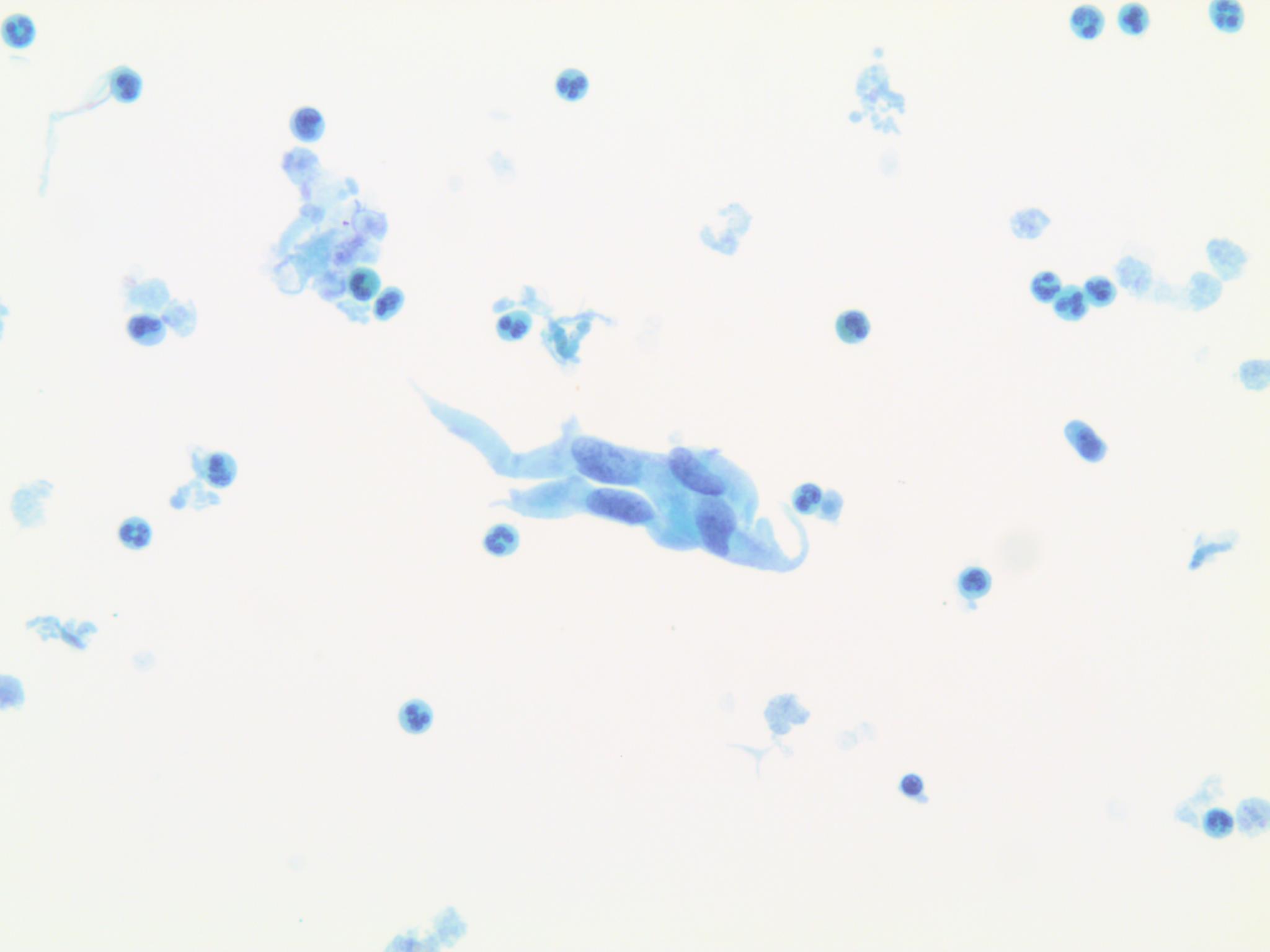
Figure 2. Liver FNA prepared on a ThinPrep, pap stain, 20x: Image chosen to illustrate the overall acute inflammation seen in a 20x field as well as the obvious spindle cell shaped cytoplasm and oval or cigar shaped nuclei.
Figure 2. Liver FNA prepared on a ThinPrep, pap stain, 20x: Image chosen to illustrate the overall acute inflammation seen in a 20x field as well as the obvious spindle cell shaped cytoplasm and oval or cigar shaped nuclei.