Jump to navigation
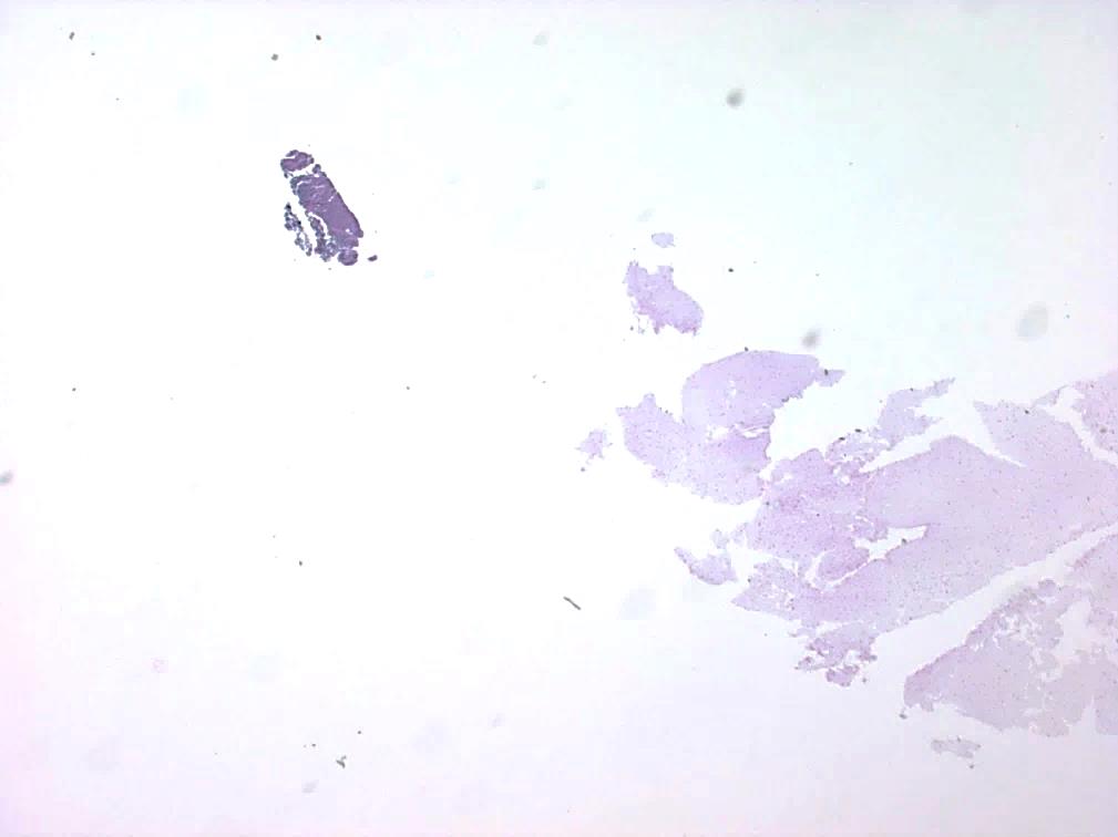
Separate fragment of tissue identified in H&E stained cell block sections.
You are not yet complete for this activity.
If you are attending a virtual event or viewing video content, you must meet the minimum participation requirement to proceed.
When the virtual event or video content is complete, please press "Next" again.
If you think this message was received in error, please contact an administrator.